X-Ray Sydney
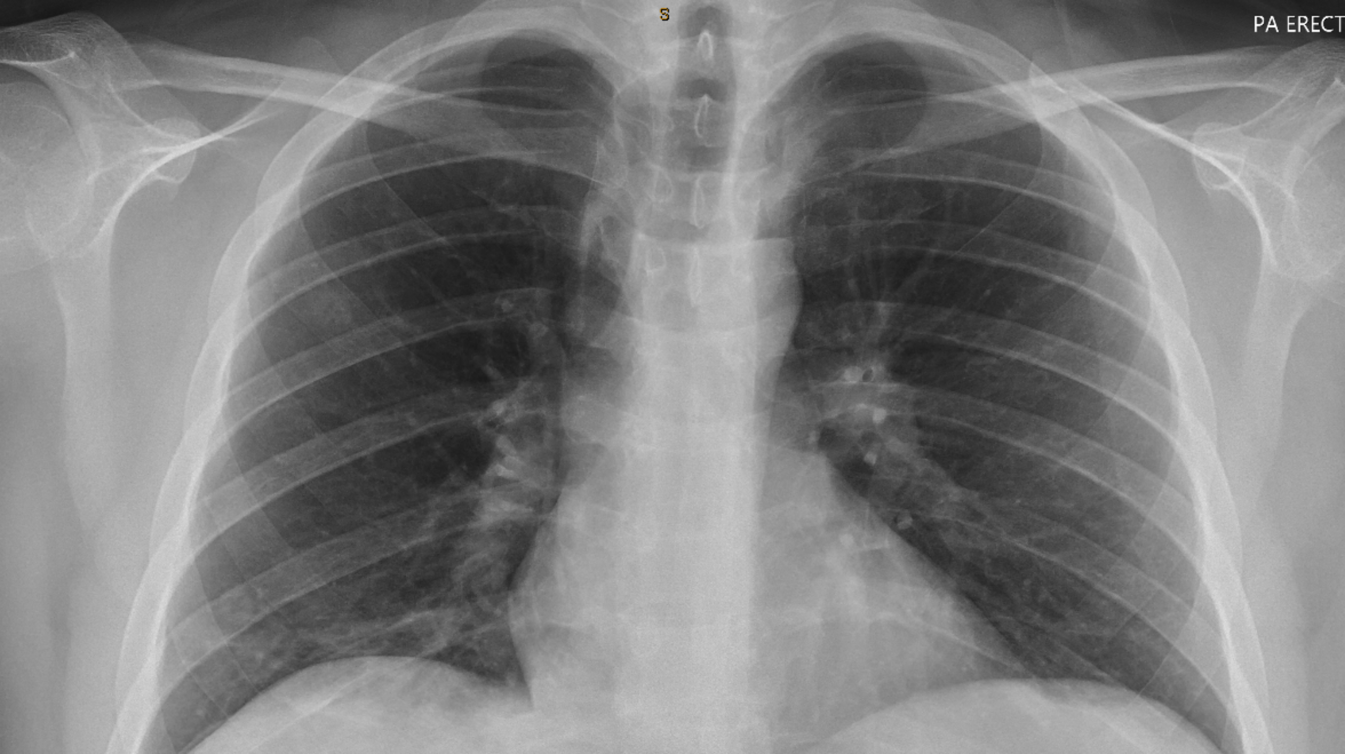
Harbour Radiology provides bulk-billed X-Ray in Sydney. X-rays are a type of medical imaging that uses a very small amount of ionising electromagnetic radiation to image the internal structures of the body. X-rays are one of the most common forms of diagnostic imaging used around the world.
We offer a bulk-billed walk-in service for X-Rays at any of our practices with an eligible referral.

Walk-in X-Ray Services Across Sydney
All X-Ray services at Harbour Radiology are bulk billed with an eligible referral and do not require an appointment. You can walk in for any X-Ray but please note you do require a doctors referral. Walk in X-Ray services include:
- Chest X-Ray
- Abdomen X-Ray
- Musculoskeletal X-Ray (hands, wrists, legs, hips etc)
- Spine X-Ray
We offer quick X-Ray scanning times averaging between 5-10 minutes and our expert Radiographers position you to take a quality image and reduce your radiation exposure.

X-Ray Common Uses and Diagnostic Procedure
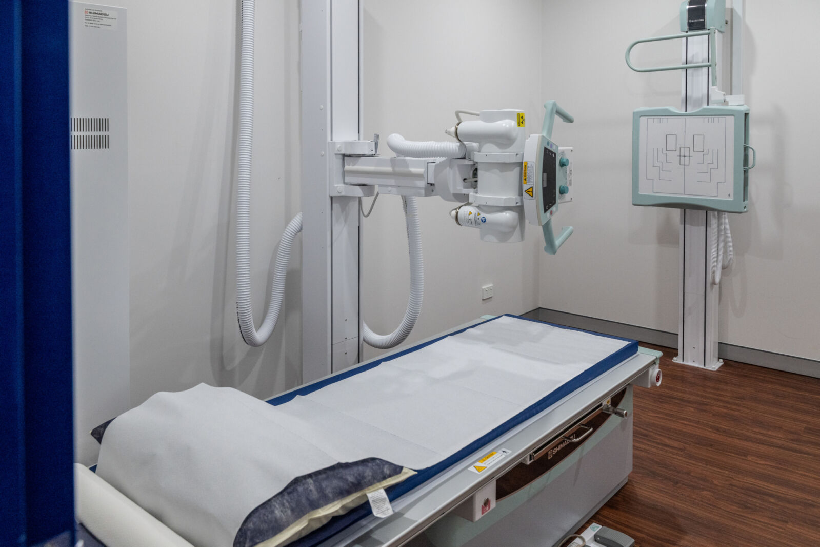
During a diagnostic X-ray procedure, the patient is positioned between an X-ray machine and a special film or detector. The X-ray machine emits a controlled beam of X-rays that pass through the body. Different tissues and structures in the body absorb or block varying amounts of X-rays. This results in the creation of a two-dimensional image that shows the internal structures, such as bones, organs, and tissues.
X-Ray imaging is the world's most common medical imaging scan. It is utilised in and out of hospital, for a wide range of diagnostic purposes, including:
- Assessing or diagnosing fractures
- Checking for foreign bodies (such as ingested coins)
- Assessing the lungs
- Post surgical alignment of bones
X-Ray imaging is one of the fastest modalities, with some scans taking less than a minute to complete. The images are immediately available to your referring practitioner, our team of Radiologists reports on every X-Ray to provide an expertly trained diagnosis of each scan.
Frequently Asked Questions
What happens during X-Ray examinations?
During an X-ray examination, the process typically involves the following steps:
- Preparation: The radiographer, a trained X-ray technologist, will call your name and lead you to an examination room. They will explain the procedure and prepare you accordingly. Depending on which part of your body is being examined, you may be asked to stand, sit, or lie down.
- Positioning: You'll be positioned appropriately for the X-ray. This positioning ensures that the area of interest is properly aligned with the X-ray machine to obtain clear images. It's crucial to stay still during the procedure to avoid blurring the images.
- X-ray Imaging: The X-ray machine emits a controlled beam of X-rays, which passes through your body and onto a special film or detector. Different tissues and structures in your body absorb or block varying amounts of X-rays, creating a shadow image of the internal structures.
- Image Processing: After the X-rays are taken, the radiographer processes each image to ensure quality. This may involve adjustments to enhance visibility or clarity. Sometimes, additional images may be needed to capture different angles or provide more information.
- Evaluation: Once the images are processed, a radiologist, who is a specialized doctor trained in interpreting medical images, carefully evaluates them. The radiologist looks for abnormalities, injuries, or other signs of medical conditions.
- Diagnosis and Reporting: Based on their evaluation, the radiologist makes a diagnosis and produces a written report detailing their findings. This report is then sent to your referring doctor, who will discuss the results with you and determine the appropriate course of action.
Throughout the examination, the process is typically straightforward, and you shouldn't feel any discomfort. You're encouraged to ask questions at any stage if you have concerns or need clarification.
How do I prepare for a plain X-Ray?
Preparing for an X-ray examination is generally straightforward. Here's what you can do:
- Bring Necessary Documents: Make sure to bring the X-ray request form or referral letter from your doctor. X-ray examinations typically require a doctor's order, so it's important to have this documentation with you.
- Inform About Pregnancy: If there's any chance you may be pregnant, inform the radiographer before the examination. Patient safety, particularly for unborn children, is a top priority, and adjustments may be needed in such cases.
- Clothing and Jewelry: Be prepared to remove certain items of clothing, jewelry, or accessories that contain metal objects, as these may interfere with the quality of the X-ray images. Items like watches, necklaces, and clothing with metal zips should be removed.
- Wear Comfortable Clothing: Since you may be asked to change into a gown for the examination, wearing comfortable clothing that is easy to remove and put back on can be helpful.
- Follow Instructions: Listen carefully to any instructions given by the radiographer or healthcare staff regarding positioning and staying still during the X-ray procedure. Cooperation helps ensure the quality of the images obtained.
- Relax: X-ray examinations are generally quick and painless. Try to relax during the procedure to facilitate accurate imaging.
By following these simple steps, you can help ensure a smooth and successful X-ray examination. If you have any questions or concerns about the procedure, don't hesitate to ask the healthcare staff for clarification.
Do you accept all X-Ray referrals?
Harbour Radiology accepts all referrals for X-ray in Sydney. The referral does not have to be written on a Harbour Radiology referral form in order to be accepted - we accept all referrals from all medical centres.
Will my X-Ray be bulk-billed?
Harbour Radiology bulk bills all X-ray examinations in all our locations (provided that you are a Medicare card holder and have a referral by a medical practitioner).
Do I need to make an appointment for X-ray examination?
We recommend that patients to make an appointment for X-Rays to avoid waiting times however walk-in patients are also accepted. Please note the wait times does vary depending on how many patients are present on the day. In most cases we can do the x-ray as soon as you attend the practice.
How do I get my X-Ray results?
Your doctor will receive the images and a written report which is usually sent electronically.
Our doctors will communicate the urgent findings with your doctor.
You can see the results and images online on your patient portal.
It is important that you discuss the results with the doctor whom referred you.